

The operation of closed or open reduction of the dysplastic force of the infant or child is extremely difficult, requires experience, in an area where in a small space, there is a disturbance in the anatomy and the approach to the true acetabulum is difficult.
The reduction can now also be performed arthroscopically in experienced Pediatric-Orthopaedic hands.
The techniques and complications of the treatment in detail:
Treatment follows the rule, to firmly retract the head into the acetabulum, to remain in the acetabulum, and to establish normal bony coverage from the acetabulum.
Treatment is usually conservative. In dysplastic hips, where the head can be adducted (Graf 2 or 3), timely placement of a dynamic hip abduction guard (Pavlik) with systematic monitoring of good adduction, achieves rehabilitation. A good retraction of the head is required and in the absence of signs of instability, the head must be repositioned in a range of lead that does not exceed 75m of lead.
Accurate ultrasound examination of the hip in abduction is technically challenging and requires extensive experience. Radiological confirmation of good retraction ensures better insight into the treatment we provide.
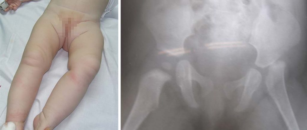
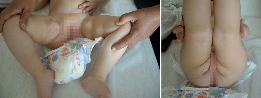
In unstable hips, management is by closed reduction after anesthesia of the infant, and precise radiographic guidance. Arthrogram control is particularly helpful and provides an accurate record of accuracy in reduction and the presence of obstacles to accurate hip reduction.
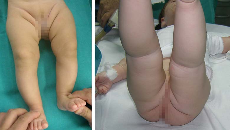
If accurate hip reduction is not achieved, or hip instability is significant and reduction is difficult and only achieved with great hip abduction, it is best to avoid closed reduction. We perform a closed transection of the adductors, which is possible to allow the reduction of the hip. If reduction in the safe abduction zone is not achieved, we perform an open reduction of the hip.
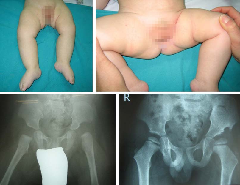
Treatment depends on the degree of deformity and the possibility of closed reduction. A closed retraction attempt is made under general anesthesia and the stability of the retraction is decided with the help of the arthrogram. Only with good head centering do we accept closed retraction, otherwise we plan open retraction, which is more likely the closer the child gets to one year old.
It is possible that after closed reduction and immobilization, if good head position is not maintained or there is insufficient bony coverage of the head, open reduction or femoral or acetabular osteotomy or a combination may follow, depending on the radiographic picture of the dysplastic hip.
Closed reduction can be attempted beyond the age of one year, but as we approach walking age and move away from the age of one year, the probability of successful closed reduction becomes smaller.
Treatment can also be performed arthroscopically. The hip is then immobilized in a hip spica plaster bandage for a period of 6-8 weeks, followed by prophylaxis with guardians for the time required for the proper coverage of the head by the bone rim of the acetabulum.
In case of bilateral dislocation of the hip, although it is possible to do the operation at the same time, it is considered safer to deal with the hips at different times, the reduction of the second hip is done towards the end of the application of the hip spica of the first hip.
As the child grows and moves away from the age of one-year, open reduction becomes the treatment of choice.
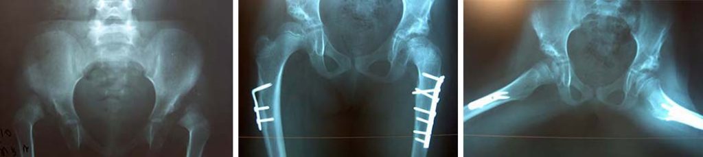
In cases of high dislocation, a combination of open reduction, femoral and pelvic osteotomy is needed, a combination that ensures the best possible reduction. The older the child, the technical difficulties of the operation increase.
At older ages, shortening of the femur is needed to achieve hip reduction. In unilateral hip dislocation, this major operation must be performed to improve walking, while at the end of childhood, bilateral hip dislocation raises the question of whether it should be treated surgically. It is undoubtedly a surgical challenge, but the end result is not always what was initially planned.
Closed reduction can be attempted beyond the age of one year, but as we approach walking age and move away from the age of one year, the probability of successful closed reduction becomes less.
In unstable hips, management is by closed reduction after anesthesia of the infant, and precise radiographic guidance. Arthrogram control is particularly helpful and provides an accurate record of accuracy in reduction and the presence of obstacles to accurate hip reduction.
In dysplastic hips, where the head can be adducted, early placement of a dynamic hip abduction guard (Pavlik) with systematic monitoring of good adduction achieves rehabilitation.
In high dislocations or failure of conservative treatment, open reduction follows with modern osteotomy of the femur or pelvis when stabilization is needed.
Next is the head retraction stability check. If abduction and medial rotation beyond 70m is needed, hip rotation and hip flexion osteotomy is planned, which is done at the same surgical time. If there is instability in hip extension because the head is uncovered in its anterior upper part, a Salter pelvic osteotomy is performed, which stabilizes the hip dislocation.
This operation is extremely difficult, requires experience, in an area where in a small space, there is a disturbance in the anatomy and the approach to the true acetabulum is difficult.
The hip is then immobilized in a hip spica plaster bandage for a period of 6-8 weeks, followed by prophylaxis with guardians for the time required for the proper coverage of the head by the bone rim of the acetabulum.
Dealing with such a serious problem can lead to complications. These are related not only to the condition itself but also to the degree and results of medical interventions.
Recurrence of the dislocation, insufficient reduction, persistence of acetabular dysplasia, occurrence of ischemic necrosis of the femoral head, short neck, leg length discrepancy, limping are among the possible complications.