

Ultrasound examination of the neonatal hip
Ultrasound and radiological examination of neonatal hip dysplasia
Some newborns, during the first days of life, show indications of developmental hip dysplasia, most of which become normal in the first 2-3 weeks of life, without a pathological finding. Even though such an event is common, hip development requires clinical and ultrasound monitoring.
The first objective recording of the neonatal – infant hip is done with the ultrasound examination, which is the predominant examination of choice.
Ultrasound is the safest method for objectively recording any pathology related to the infant hip, especially before the appearance of ossification nuclei. We avoid unnecessary radiological examination of normal infants.
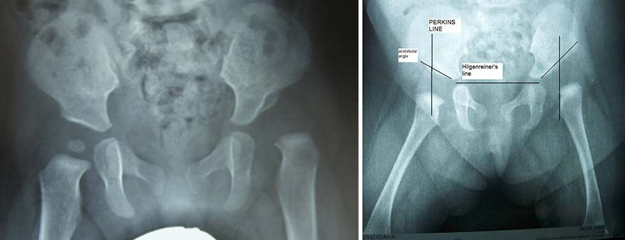
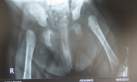
In the radiological examination we evaluate the symmetry of the pelvis. The diagnosis of dysplasia is easier when ossification nuclei have appeared.
Computed tomography and magnetic resonance imaging (MRI) scans provide information regarding the hip morphology and are primarily used for accurate hip repositioning in the hip spica.
Ultrasound examination of the neonatal hip
Ultrasound provides accurate information on the anatomic elements of the hip and is undoubtedly the safest method for recording the pathology of the infant hip, especially before the appearance of ossification nuclei. This method avoids the unnecessary radiological examination of normal infants.
The first 6 to 8 weeks after birth is the most reliable period for accurate examination. In the immediate neonatal period, the increased cartilage ratio and elasticity, can lead to misdiagnosis -usually type 2- and overtreatment of normal hips.
Radiological examination of the neonatal hip
The diagnosis of hip dysplasia after clinical and ultrasound examination, should be followed by radiological examination.
Radiological examination during infancy and early childhood evaluates various parameters. The clinician can measure the acetabular angle, the percentage of head coverage and the CE angle (the angle formed by the perpendicular line passing through the center of the head and its line formed by the union of the center of the head with the upper outer edge of the acetabulum), which are indicators mainly used during the treatment of dysplastic hip. (Severin classification).
Computed tomography and magnetic resonance imaging (MRI) scans provide information about the hip morphology, and are primarily used to evaluate the exact position of the hip in the hip spica.
It is very important, during both the ultrasound and radiological examination, to position and immobilize the baby correctly, in order to avoid the inclination of the pelvis, leading to inaccurate conclusions.
Arthrography of the neonatal hip
Arthrography is a very reliable record in the treatment of dysplastic hip. During the examination a small amount of X-ray fluid is injected, which allows the visualization of the hip joint.
In hip dysplasia, the radiolucency stagnates the area of the fibrous part of the acetabulum and the position outside the acetabulum head is presented, the pathological image of the labrum. It is possible to record the hourglass image of the bursa from the impression created due to the pressure of the psoas muscle.
We present the repositioning of the hip, using specific repositioning manipulations to control the new placement of the repositioned head and the coverage of the bony and cartilaginous part of the acetabulum.